
Microscopy and Imaging Shared Resource (MISR)
Services and Instrumentation
Services offered by the Microscopy and Imaging Shared Resource include:
- Consultation on experimental design and the choice of the most appropriate imaging modalities and systems.
- Initial and ongoing training on the various imaging platforms
- Assistance with the acquisition of images and experimental troubleshooting
- Training and assistance with the use of image analysis software and the development of custom analysis approaches
- Acquisition and processing of data on some systems.
- Molecular cytogenetics service
- Help preparing the relevant sections of grant submissions and manuscripts
- Supply optimal tissue culture ware and other consumables for pilot projects
- Supply a range of validated imaging reagents and DNA constructs
- Periodic lectures and workshops on a range of imaging and analysis topic
MISR Instruments and Key Features (DC campus)
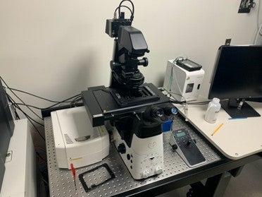
Leica SP8AOBS++. Laser scanning confocal microscope with 10 laser lines (405, 442, 458, 476, 488, 496, 514, 561, 594, 633nm), 8 channel ABOS, 8 Channel AOTF, 5 channel spectrometer (400-800nm/5nm), 3 HyD photon counting detectors, 2 Hamamatsu PMTs, bright-field detector with DIC, tandem scanner (12KHz TCS, 24 KHz resonance), closed loop auto focus, scanning stage (20X40nm R, 1um A), a range of objectives including 10x/0.4, 20x/0.75 W/G/O, 40x/1.03 O, 40x/1.1 W, 63x/1.4 O, a full chamber with heat/CO2, full range of LAS software modules to facilitate FRE, FRAP, multi-point time-lapse confocal imaging, etc.
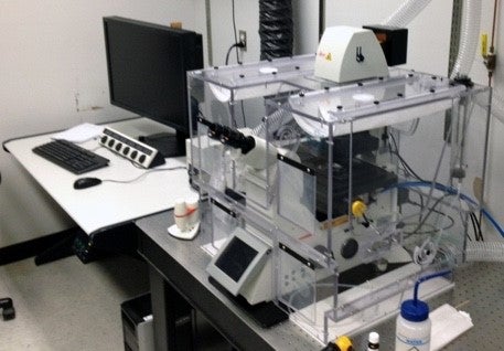
Olympus FVMPE-RS with DIVER detector: Custom imaging system for multiphoton and fluorescence lifetime imaging. Built on an Olympus FVMPE-RS upright frame equipped with Insight x3 (700nm – 1300nm pulsed) and DPSS 405nm lasers, a range of objectives (including 10x/0.04 A, 20x/0.7 A, 25x/1.05W, 40x/0.8 W), galvo (pixle time 2ms – 1S) and resonant (30-438 fps) scanners, 2 cooled GaAsp-PMTs + multi-alkali photomultipliers, DIVER detector and FLIMBox (ISS) for deep imaging, NADH/FAD imaging, second and third harmonic generation FLIM and Phasor based fluorescence lifetime imaging/analysis, and an array of acquisition and analysis packages to support available imaging modalities and for multiple area time-lapse imaging (MALT), metabolic analysis, etc.
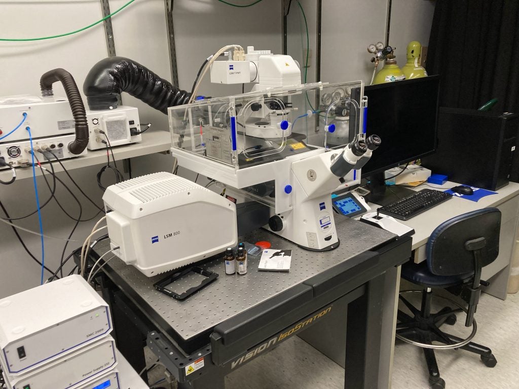
Zeiss LSM 800: Laser scanning confocal microscope designed primarily to support wide-area confocal time-lapse imaging. The system which is housed in an environmental chamber with controlled temperature, humidity and CO2 leves, is equipped with 4 laser lines (405, 488, 568, 647nm), objectives (including 10x/0.3, 10x/0.45, 20x/0.8, 63x/1.4 O, 63x/1.2 W), three GaAsP detectors including a super-resolution Airyscan detector, coded motorized stage (130×100 step), Colibri 7 solid-state light source and AxioCam 506 monochrome camera to facilitate widefield fluorescence imaging in addition to confocal super-resolution modes.
Abberior STEDYCON: Based on a Nikon Ti2-E inverse body, the Abberior STEDYCON super-compact Nanoscope provides access to STED-based super-resolution microscopy (<40nm R) as well as LS confocal microscopy and fluorescence lifetime imaging (FLIM) capabilities. The system is equipped with 4 laser lines (405nm – cw, and 488nm, 561nm, 640nm pulsed), and a STED laser (775nm pulsed), a 100X 1.45NA STED oil objective and 20X air objective for ROI identification, two STED and four confocal imaging channels with 4 single photon counting APD modules, providing three detection bands (500–550nm, 580-630nm, and 650-700nm). The Picoquant TimeHarp imaging hardware provides 25ps time resolution with a sustained count rate of 40MHz, with a synchronization unit between the Picoquant and Abberior hardware. The stage is equipped with an incubator chamber and stage-top z-piezo for z-drift compensation.
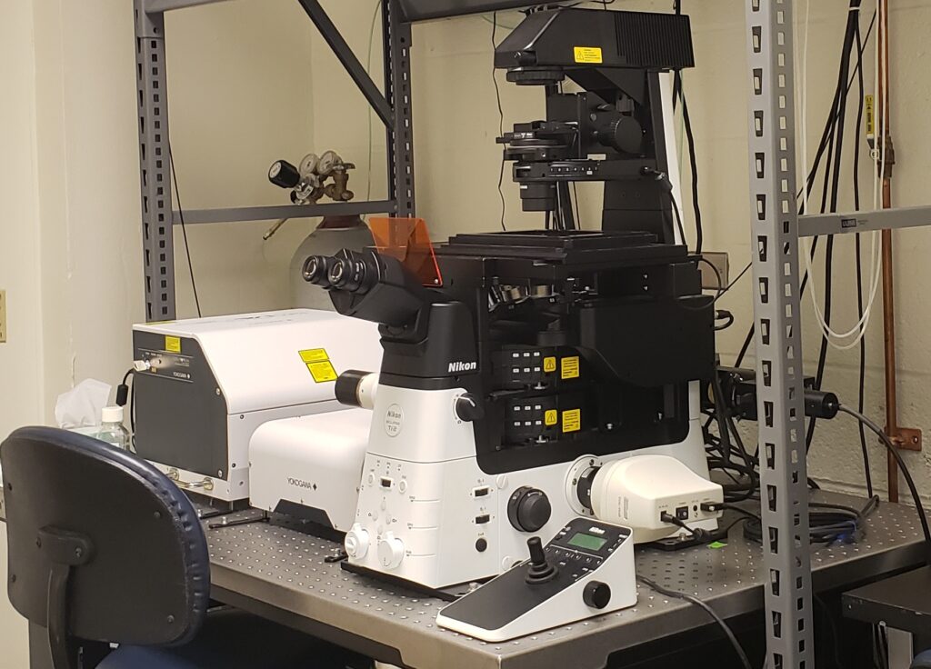
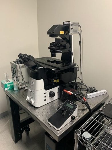
Nikon CSU-W1 Spinning Disk Confocal with SoRa super-resolution and TIRF: Based on a fully motorized Nikon Ti2E inverted microscope, this system is equipped with a Yokogawa CSU-W1 dual disk, dual camera SoRa Confocal Scanner with lines at 405nm, 488nm, 561nm, and 640nm, with a separate launcher for TIRF imaging with lines at 488nm, 561, nm, and 640nm. Confocal resolution is ~250 nm, SoRa ~ 100 nm, and Z resolution in TIRF is ~120 nm. A range of high NA objectives provide magnifications of 4X, 10X, 20X, 25X, 40X, 60X and 100X. A 405nm laser allows stimulation and photobleaching and the system is equipped with a stage-top incubator of t facilitate live cell imaging. Both spinning disk/SoRa and TIRF are fully automated through Nikon elements, and a separate workstation allows off-line processing and analysis with NIS-Elements and other software.
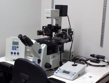
Olympus IX71 Inverted Epi-Fluorescent Microscope: The Olympus IX71 epi-fluorescent microscope is a “drop in” wide field fluorescence system for investigators who need quick access to a simple imaging platform to evaluate samples by brightfield and/or fluorescence, to determine transfection efficiency etc. The instrument is equipped with a range of objectives (including 10x/0.30, 20x/0.40, 40x/0.60), and filters for DAPI/Hoechst, FITC, TRITC.
Nikon SMZ-1500 Fluorescent Stereoscope: The SMZ-1500 stereoscope allows the stereoscopic visualization of tissues in brightfield using reflected or transmitted light, pseudo darkfield imaging, and by epi-fluorescence. Objectives available include 0.5x (0.375x – 5.6x), 1.0x (0.75x – 11.25x), and 1.6x (1.6x – 18x), and filters are installed for GFP (Ex 480nm/40nm, Di 505nm, Em 535nm/50nm), DsRed (Ex 545nm, Di 570nm, Em 620nm), and Q-dots ( Ex 460nm, Di 475nm, Em 655nm).
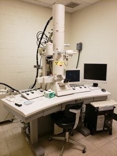
Hitachi H7600 Transmission Electron Microscope: The Hitachi H7600 transmission electron microscope has an accelerating voltage of 40-120KV, and is capable of a low magnification range of 50-1000x and in high magnification mode of 700-60,000x, and is equipped with a CCD camera for image acquisition. The instrument is supported by a dedicated laboratory space equipped with the necessary ancillary equipment including a Reichert Ultracut E ultramicrotome, and a dedicated chemical fume hood. Investigators must work with the MISR manager to use this instrument.
Olympus IX71 – Eppendorf FemtoJet Microinjection System: The dedicated micro-injection system consists of an Olympus 1X71 inverted microscope equipped with 10x/0.3 and 20x/0.4 objectives for brightfield and Normaski imaging and an Eppendorf Femtojet holding and micro-injection system. This platform allows the microinjection of adherent or suspension cells and is idea for pro-nuclear injection of mouse embryos and the manipulation/injection of blastocysts.
MISR Instruments and Key Features (NJ campus)
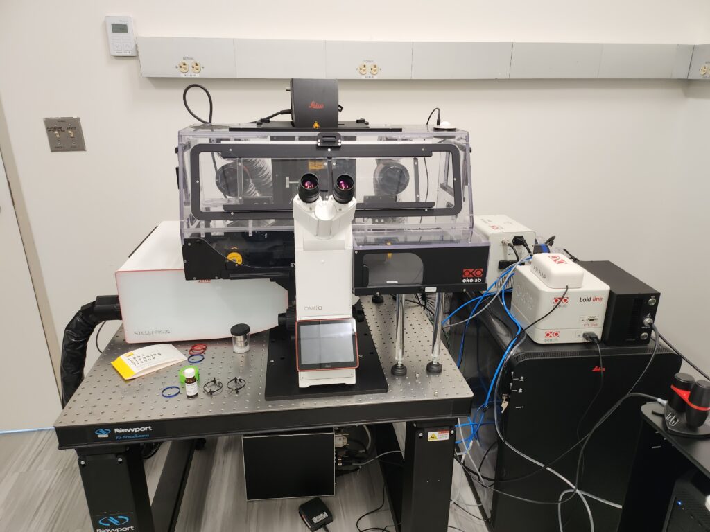
Leica Stellaris 5 Inverted Confocal: Equipped with a Humidity and CO2 controlled environmental chamber, laser lines at 405nm, 488nm, 561nm, and 638nm, and 10X, 20X, 40X, and 63X objectives, an 8kHz tandem scanner, and 4 power HyD S detectors, this system is ideal for time-lapse, live-cell conical imaging all controlled by the LAS X Stellaris control software.
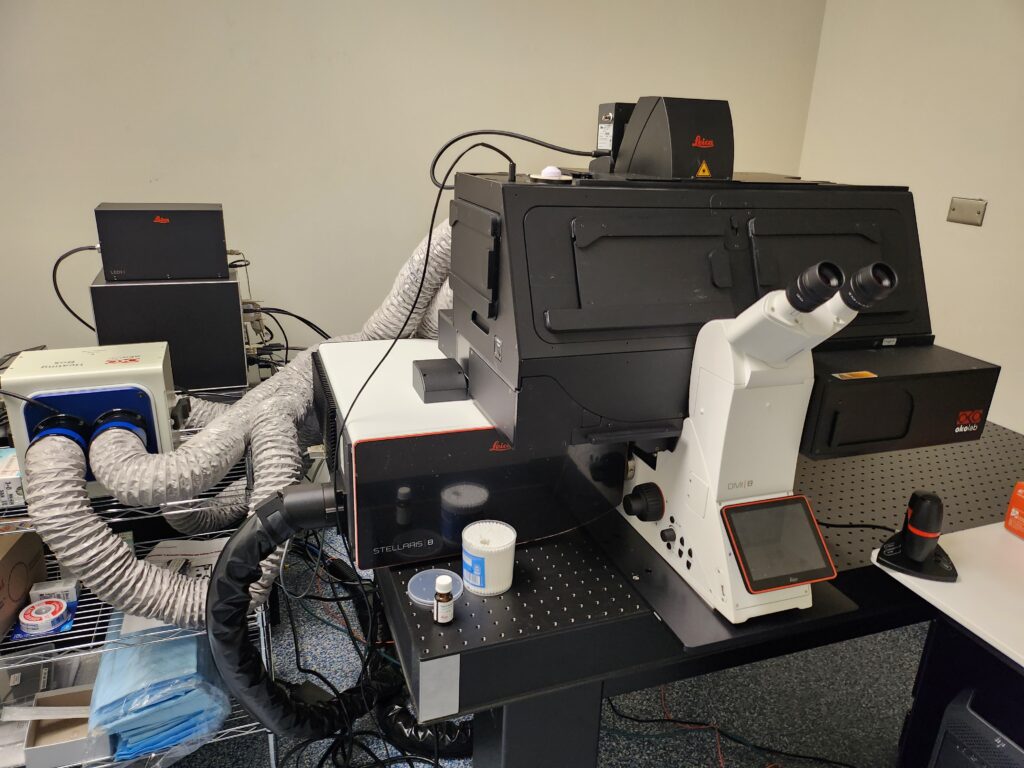
Leica Stellaris 8 Inverted Confocal: This system, equipped with a Humidity and CO2 controlled environmental chamber, laser lines at 405nm, 488nm, 561nm, and 638nm, and 10X, and 25X, objectives, FOV scanner and 4 power HyD S detectors, is optimized for intra-vital conical imaging all controlled by the LAS X Stellaris control software.
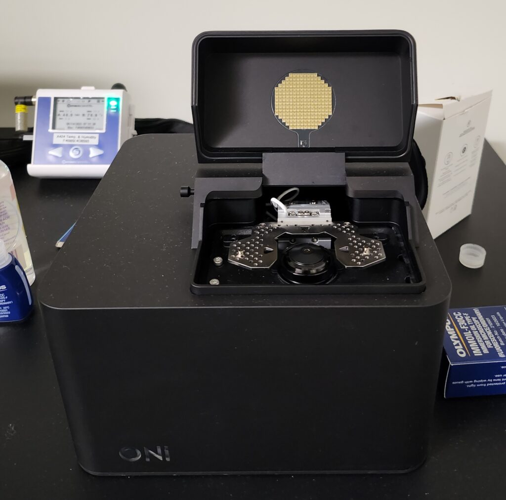
ONI Nanoimager Super Resolution Microscope: The ONI Nanoimager a PALM / dSTORM based super-resolution imaging platform that is also capable of TIRM, epifluorescence and smFRET imaging. it is equipped with a 100X 1.45NA oil immersion objective and has laser lines at 405nm, 488nm, 561nm, and 640nm. This system is optimized for single-molecule localization and astigmatism adjusted 3D imaging.
Nikon Ti2 A1R LSM Confocal Microscope (NJ): The Nikon Ti2 microscope equipped with an A1R LSM confocal unit and a Tokai Hit incubation chamber is ideal for imaging both fixed and live cell samples. The instrument has laser lines for DAPI, FITC, TRITC, Alexa-647 (405, 445, 488, 514, 561, 640nm) and a range of objectives (4x, 10x/0.45 A, 20x/0.75 A, 40x/0.95 A, 40x/1.3 O, 60x/1.4 O).
Nikon Ti2 widefield Epi-Fluorescence microscope (NJ): The Nikon Ti2 epi-fluorescent microscope is equipped with a range of objectives (4x, 10x, 20x, 40x, 60x O, 100x O), and filters for DAPI, FITC, TRITC, Cy5, CFP, YFP, mCherry, and a Tokai Hit incubation chamber. The system is ideal for brightfield and widefield fluorescent imaging of fixed and live-cell specimens with images captured with a Hamamatsu camera.
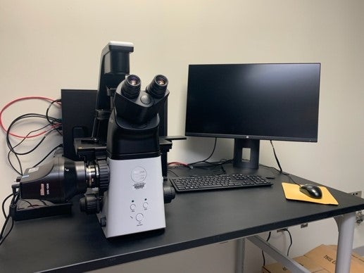
Nikon Ti2R with DS-Qi2 color camera (NJ): The Nikon Ti2R epi-fluorescent platform is equipped with a range of objectives (4x, 10x, 20X, 40x, 100x O), and filters for DAPI, FITC, TRITC, and Cy5. The color Ds-Qi2 camera makes this platform ideal for brightfield and fluorescent imaging of a range of samples.
Image Analysis Workstations
Multiple stand-alone image-processing workstations are available on the DC and NJ campuses. These workstations are equipped with off-line versions of a variety of image acquisition/analysis packages including NIS-Elements, MetaMorph, Volocity , Huygens, ImageJ with custom plugins etc.